Dr Elisabeth Kugler | Director of Zeeks – Art for Geeks Ltd | Twitter @Zeeks_art
| Instagram @zeeks.artforgeeks | Facebook Zeeks – Art for Geeks
Zebrafish?
Yes, zebrafish. The ones you can buy in the pet shop!
Zebrafish are important models in biomedical science to answer fundamental questions of body development, health, and disease. A lot of work is focused on understanding how tissues and organs develop, because once we understand how something is formed, it is easier to understand how it is maintained in health and how it is breaking down in disease.
While adult zebrafish are about 2-3 centimetres in size (which is the length of a matchstick), when studying developmental processes many labs examine zebrafish embryos (smaller than the tip of a matchstick). Thus, we need microscopy to elucidate and reveal the hidden microscopic worlds.
The microscope
While the size of the embryos seems very small, in microscopic terms this is actually very large and we normally only study tissues in the scale of a few micrometers rather than hundreds of them. This poses a challenge as light interacts with matter [1]. To address this, light-sheet microscopy (LSFM) has become the ideal solution to acquire data in large tissue samples.
In a nutshell, LSFM uses a sheet of light to illuminate a thin optical section of a sample and the data are acquired by an uncoupled emission detection objective. This means, that while in conventional confocal microscopy, a whole cone of light shines through the sample, in LSFM it is a thin plane that is illuminated and this whole plane is imaged at once. Together, this results in less photobleaching, fast acquisition, and large fields-of-view [2].
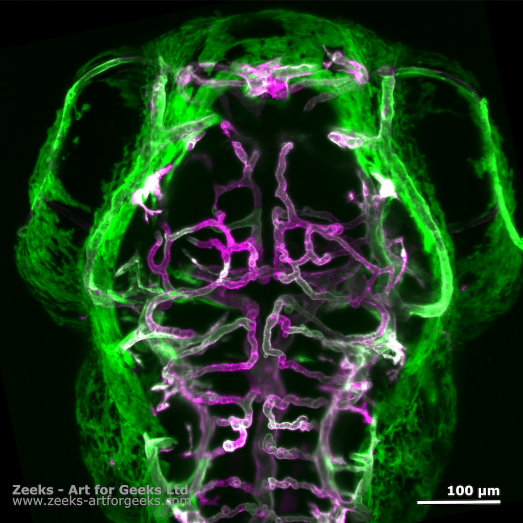
The science art
As the acquired data are rather large in microscopy terms, this means that there is a lot of data to work with for analytical purposes, but also to endeavour on data examination paths for science art. Dr Elisabeth Kugler (Twitter @KuglerElisabeth) has used her microscopy data in the last few years to share her scientific passion using visual science art [3].
More recently, Elisabeth has formed the company Zeeks – Art for Geeks Ltd, which is all about data. This will allow her to focus on all things data and explore new avenues how to communicate scientific research using science art.
References
[1] E. Kugler and E. Reynaud, “LSFM series – Surfing on the data freak wave! Part II: Before imaging: Know your sample (geometry),” FocalPlane, Oct. 10, 2020. https://focalplane.biologists.com/2020/10/10/lsfm-series-surfing-on-the-data-freak-wave-part-ii-before-imaging-know-your-sample-geometry/ (accessed Nov. 16, 2022).
[2] J. Huisken, J. Swoger, F. Del Bene, J. Wittbrodt, and E. H. K. Stelzer, “Optical sectioning deep inside live embryos by selective plane illumination microscopy,” Science, vol. 305, no. 5686, pp. 1007–1009, Aug. 2004, doi: 10.1126/science.1100035.
[3] the Node, “SciArt profiles: Elisabeth Kugler,” the Node, Jul. 28, 2021. https://thenode.biologists.com/sciart-profiles-elisabeth-kugler/science-art/ (accessed Nov. 16, 2022).


